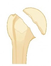
Simple fractures

We will describe fractures as ‘simple’ when they have two fragments, as opposed to complex fractures (which combine several types of simple fractures at the same time).

A simple fracture can separate the tuberosities or cervix from the humerus.

The challenge of tuberosities is the function of the cap

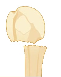
For fractures of the cervix, in addition to the displacement limiting the good function of the shoulder, there is a risk of necrosis of the humeral head and therefore of sagging of the latter with risk of early osteoarthritis and rapidly progressive (indication of shoulder prosthesis )

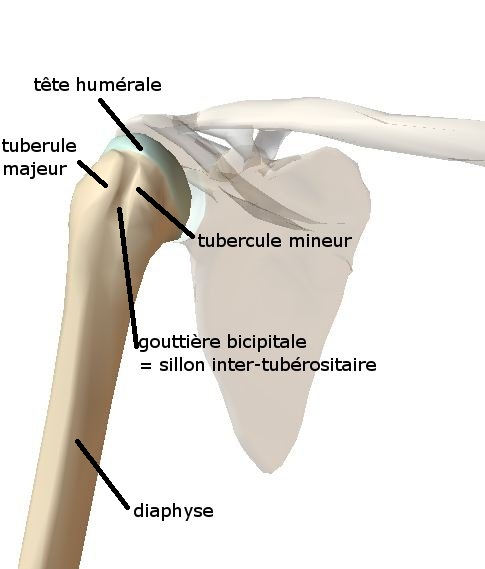
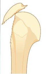
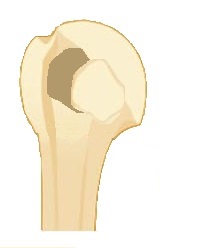
Fractures of tuberosities

Very frequent, they occur during a fall or accompany a shoulder dislocation (tearing of the tuber).

The fragment can be minimal (bone impaction or crushing) see take the whole massif.

Their displacement is due to the action of the supraspinatus and / or infraspinatus tendon (ascension of the fragment and / or posterior retraction).

Not exceptionally, the fracture is sometimes undiagnosed on initial radiographs, this is a common reason for frequent consultation, especially during the winter sports season.

In the majority of cases there is no displacement, the immobilization of the shoulder by jar or abduction cushion (with neutral rotation if infra-spinous concerned) is envisaged during 4 to 6 weeks. The suites are often very long at around 6 months or even more, because of an associated contusion of the tendon inserted on it and the fact that tendon traction is done on a weakened and convalescent bone).

They must be fixed (osteosynthesized) if the displacement is greater than 5 millimeters and the fragment is voluminous. Smaller fragments can be reinserted in the same way as open rotator or arthroscopic rotator cuff tendons. the sequences are identical then to a tendinous suture.

Although less frequent, the diagnosis is sometimes also difficult especially in the absence of displacement and may require a scanner or an MRI.

Due to the presence of the subscapularis tendon, the displacement is medial. The immobilization is done by jutting in internal rotation (hand on the belly).

The surgical indications are identical.

In any case, it is better to operate such a displaced fracture than to have to deal with a malunion (consolidated fracture in bad position) which can be troublesome (sub-acromial or sub-coracoidal conflict) or lead to a malfunction of the rotator cuff.

Vicious callus surgery is very complex, non-consensual and extremely random in its results, generally much less favorable than a good management of the initial fracture.

Fractures of the humeral neck

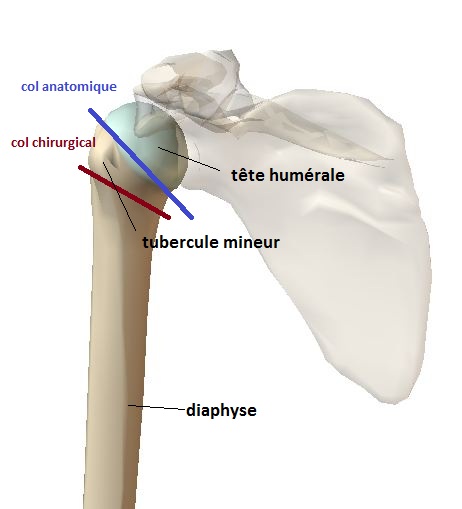
With regard to fractures of the humeral neck, it is necessary at the outset to distinguish the fractures of the surgical neck and the anatomical neck (see anatomy humerus)

By definition the most frequent, extra-articular fractures generally meshing (diaphysis is entered in the upper fragment) therefore stable (does not move under the effect of a few limited movements or through muscle tractions). They most often come from medical treatment (immobilization then physiotherapy), it often appears a bruise on the arm that migrates by gravity to the elbow (if painful may require lymphatic drainage)

In case of displacement they can be osteosynthesized according to the surgeon’s habit (nail, pins, plate). We personalize this type of fracture (often by a percutaneous technique).

Exceptionally isolated (simple fracture), most often constitutes a component of complex fractures (see chapter).

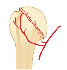
These are intra-articular fractures with the specificity of a risk of joint stiffness due to the proximity of the joint capsule and a risk of avascular necrosis [ avascular necrosis] of the humeral head due to the interruption of the vascularization to the target of the head (see diagram below vascularization called ‘terminal’, the blood borrows only one possible way without possibility of substitution …)

If there is no more connection with the joint capsule this risk is major with evolution towards the deformation of the head, pain and a very severe loss of mobility. It is then necessary to consider the replacement of the head (humeral head prosthesis) if possible (according to age) before the intervention.

It is of terminal type, that is to say that the arterial blood flow is unique and that its interruption causes the necrosis (tissue death) of the head.

The clinical result may be a weak to medium gene if necrosis affects only part of the humeral head.

In case of massive necrosis the evolution is towards a collapse of the head which no longer allows a sliding on the glenoid and therefore is at the origin of a limitation of the articular amplitudes and pains.

Replacing the head with a prosthesis is the most effective intervention.