The clavicle is a long bone, sort of upper bumper thorax protecting nerves, vessels for the upper limb but also the upper lung. The traumatology of the clavicle is frequent, we will distinguish in order of frequency:
- fractures of the middle third, often displaced, sometimes with three fragments, are the most frequent whether in adults or in children,
- fractures of the outer third, located close to the acromioclavicular joint and especially clavicualire ligaments uniting the clavicle and the scapula
- fractures of the internal third, initial radiological diagnosis sometimes difficult, near the sternum less frequent.
Fracture of the external third clavicle
For some of these fractures there is the same consequence as the acromioclavicular disjunctions, when the fracture detaches the coraco-clavicular ligaments there may be a fall of the stump of the shoulder and important displacement of the fragments between them.
in these cases the bone consolidation even in malunion can be very random and justifies for us an intervention very often acute.
Broaching can be performed, sometimes also crossing the acromion which greatly stabilizes the assembly; there is specific material often metal plates that we prefer.
Finally the same surgical attitude can be used as for acromioclavicular dislocations (see chapter) with implantation under endoscopy of an artificial ligament passed under the coracoid.
Fracture of the middle third clavicle
Orthopedic treatment
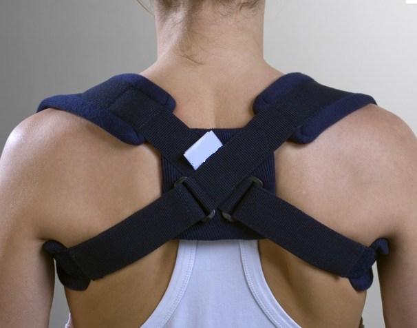
It is most often rings cloth, passed in 8 around the shoulders and squeezing in the back. They have a role first analgesic because these fractures are very painful the first time especially if they are not immobilized. These rings often allow a maintenance or even partial reduction of fragments between them, especially if there is significant overlap. fractures usually consolidate forming a malunion sometimes causing a lump that tends to diminish over time. It happens that a fracture does not consolidate and requires a surgical intervention (osteosynthesis sometimes with bone graft).
The rings should be regularly torn without creating swelling of the hand or upper limb, ants. In exceptional cases of tight rings, deep vein thrombosis (= phlebitis, the most serious risk) has been observed which requires immediate removal of the rings, emergency consultation with a doctor (Doppler ultrasound and anticoagulant injection).
Surgical treatment
Operative indications depend on the surgical team and the region (Midi-Pyrenees region little surgical attitude)
– open fracture or where a fragment is under the skin and threatens to cross it
– In case of associated lesions (fractures, nerve, vessel or organ damage) if solid immobilization is preferable or in case of non-tolerance of the rings.
– Some cases are discussed on a case by case basis:
- at the athlete of good level
- in case of very displaced fracture not improved by the rings (which the surgeon considers a high risk of progression towards a non-union).
The operative indications remained for us like most regional teams rare in front of the number of cases and especially with the good frequent evolution after immobilization.
Evolution of our indications: more surgery !!
Why ?
The orthopedic treatment (rings) does not allow to reduce correctly a very displaced fracture, the duration of complete consolidation (radiological) is frequently long (3 to 6 months or more!) with stopping sports or working at the manual worker. Non-union (= pseudarthrosis) is possible.
A consolidation in malunion can be at the origin of genes probably underestimated medically:
– a significant shortening leads to one shoulder shortens in width and one shoulder projected forward, causing compensatory pain in the shoulder or around the scapula, whose treatment is difficult …
– a bulky callus canshrink the subclavicular space (cervico-thoracic parade) and hinder the nerves and blood vessels to destiny of the upper limb.
Our current indications:
Displaced fractures in the young or active subject (sports, manual work) with:
– an inter-fragmentary space greater than 5 mm (possibly not improved by the wearing of the rings)
– a shortening greater than 10 mm (possibly not improved by the wearing of the rings) most often by overlapping fragments

Surgical techniques used:
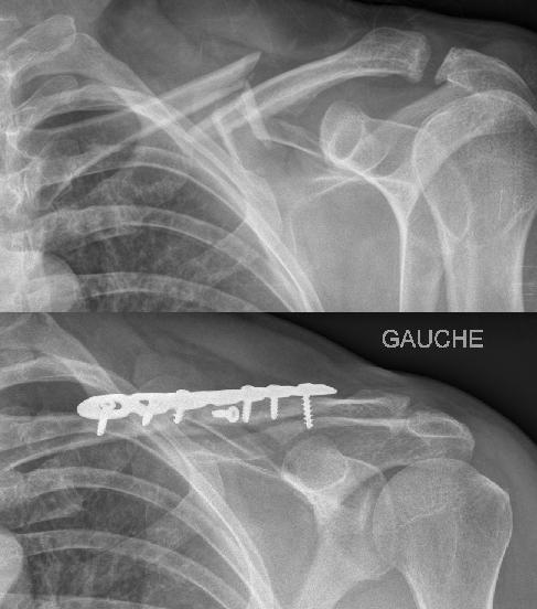
Screwed plate (see radio example above)
Most widespread technique, usable for any fracture. It requires a wide approach to position the plate and screws (3 screws on both sides of the fracture if possible). The material is often prominent, awkward under the skin (removal of frequent material)
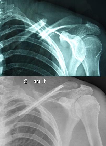
Centromedullary screw (see radio opposite)
Recent technique, being developed in France, very interesting because of the absence of material discomfort, however not applicable to all fractures (long oblique fractures, several comminuted fragments or absence of intraosseous medullary canal).
Operative suites:
Prudence 4 to 6 weeks (dujarrier or scarf), arm not always free (no driving), passive film or even self-education by limiting abduction and elevation earlier than 70 or 90 °.
Stop sports and heavy manual work until radiological consolidation
fracture of the internal third collarbone
Fractures less frequent, may be the most difficult to diagnose urgently because very little visible on X-rays.
Sterno-clavicular dislocation is the differential diagnosis, in both cases a CT scan seems essential.
Surgery remains exceptional for isolated fractures, a vascular specialist available is a necessary caution in the proximity of the large vessels of the base of the neck if an intervention is considered at a distance from the trauma.


